Aging reshapes the hypothalamus region by region, with some nuclei shrinking and others growing
Buck Institute researchers mapped hypothalamic volume and gene activity in aging and Alzheimer's, finding region-specific changes and early inflammatory shifts.
Alzheimer's disease is usually understood through the hippocampus and cortex, the brain regions where memory is organized. That framing misses something patients and families notice early: disrupted sleep, shifting appetite, and changed responses to stress. A new study from the Buck Institute for Research on Aging shows the brain structure behind those symptoms, the hypothalamus, is not a bystander in the disease. It undergoes specific, region-by-region changes in volume and gene activity that appear alongside aging and early Alzheimer's.
Why the hypothalamus matters
The hypothalamus sits at the base of the brain and orchestrates sleep-wake cycles, metabolic homeostasis, stress responses, and neuroendocrine function. Alzheimer's is more than cognitive decline; it is a systemic condition that disrupts precisely these processes. Emerging evidence points in both directions: hypothalamic dysfunction correlates with Alzheimer's pathology and may actively worsen it. Yet the region has remained understudied in human aging and disease.
The hypothalamus is not a uniform block. It is made of many specialized sub-areas, or nuclei, each with a distinct job. The suprachiasmatic nucleus, or SCN, acts as the body's master clock. The arcuate nucleus, or ARC, handles appetite and energy balance. Until now, very little was known about how these individual nuclei and their cell types change as people age or as Alzheimer's takes hold.
A two-lens approach
No single technique can answer these questions. MRI shows what is happening in living people but lacks fine resolution. Single-nucleus RNA sequencing reveals what is switched on inside each cell type, but requires actual tissue. The team, including senior authors Ashley Webb, PhD, at the Buck Institute and Manu Goyal, MD, at Washington University School of Medicine in St. Louis, built a resource called HypoAD to put the two lenses side by side. It combines high-resolution MRI from hundreds of living individuals with single-nucleus sequencing of more than 600,000 nuclei from young participants, people with Alzheimer's, and age-matched controls without dementia. The atlas is available to the research community.
Differential aging reshapes the region
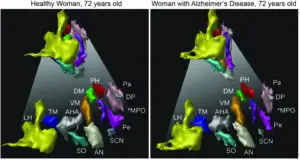
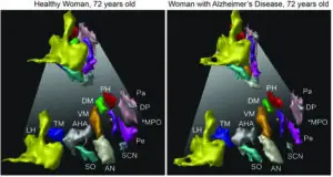
The most striking result is that the hypothalamus does not simply shrink. Measured as a whole, its volume barely changes with age. Zoom in, and the picture is structured: the lateral hypothalamus shrinks, while the SCN, arcuate, paraventricular, periventricular, and dorsomedial nuclei grow. This pattern held even after accounting for Alzheimer's status, which points to a feature of typical aging rather than the disease alone.
Differences tied specifically to Alzheimer's appeared but were not statistically significant. Compared with the effects of aging and sex, volume differences linked to amyloid-β deposition and cognitive impairment were more modest or absent. The authors suggest a more sensitive method may be needed to detect what the Alzheimer's hypothalamus does to volume.
Inflammation and the clock show up in the genes
At the molecular level, machine-learning models that separated Alzheimer's cells from control cells using gene expression alone flagged two dominant themes: inflammatory response and circadian-rhythm regulation. Microglia, the brain's resident immune cells, showed the largest number of altered genes and shifted from a resting state toward the disease-associated state described in other Alzheimer's work. The researchers frame this as a transition to an inflammatory state early in the disease, while noting their cross-sectional data cannot establish cause and effect.
Of the 254 neuron subtypes identified, inhibitory GABAergic neurons changed the most, mainly in neuropeptide and receptor signaling. One prominent group of neurons in the SCN showed disrupted vasopressin signaling, a key output of the body's clock. Because sleep and circadian problems are among the earliest signs of Alzheimer's, and sundowning, a circadian behavioral change, appears later, this gives a concrete place to start looking for why.
Total cell numbers did not appear to change. Instead, the data point to cells getting larger through increased activity of mTOR, a growth pathway, along with remodeling of material between cells and inflammatory activation of glia. The authors present this as a hypothesis for future work.
What it means, and what comes next
The findings also come with clear limits. The autopsy cohort of 14 donors is small, which is typical for this kind of study. The MRI and sequencing data come from different people, so they are compared side by side rather than combined statistically. And because the design is cross-sectional, the results show associations, not cause and effect.
For readers managing their own health or a family member's, the practical takeaway is that sleep, daily rhythms, appetite, and stress responses are part of the Alzheimer's picture, not just background noise, and that changes in these areas can appear alongside or even before memory loss. That is information worth bringing to a doctor when symptoms show up. This is a starting point rather than a final answer. The team's next step is to follow these changes over time and test whether protecting the brain's clock, and neuropeptide signaling in particular, can slow the disease or help manage symptoms. If circadian disruption is among the first things to go wrong, it may also be one of the first places where intervention is possible.
The study, "Hypothalamic volume and cell-type changes in aging and Alzheimer's disease," was published in Nature. You can read the full report here.